


 Foundational characteristics of cancer include proliferation, angiogenesis, migration, evasion of apoptosis, and cellular immortality. Find key markers for these cellular processes and antibodies to detect them.
 The SUMOplot™ Analysis Program predicts and scores sumoylation sites in your protein. SUMOylation is a post-translational modification involved in various cellular processes, such as nuclear-cytosolic transport, transcriptional regulation, apoptosis, protein stability, response to stress, and progression through the cell cycle.
 The Autophagy Receptor Motif Plotter predicts and scores autophagy receptor binding sites in your protein. Identifying proteins connected to this pathway is critical to understanding the role of autophagy in physiological as well as pathological processes such as development, differentiation, neurodegenerative diseases, stress, infection, and cancer.
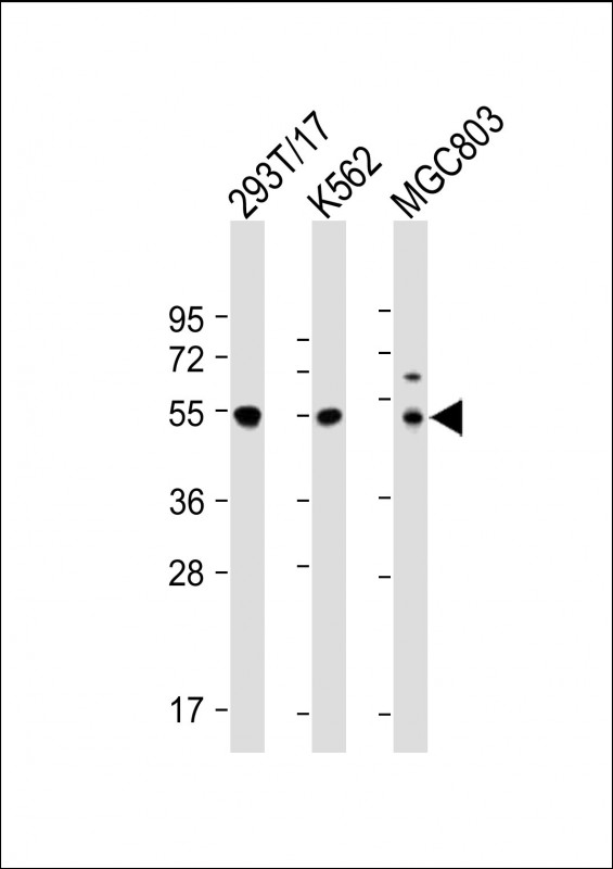
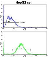
Cyclin A (CCNA2) Antibody (N-term)
Purified Rabbit Polyclonal Antibody (Pab)

- SPECIFICATION
- CITATIONS: 2
- PROTOCOLS
- BACKGROUND

Application
| FC, IHC-P, WB, E |
|---|---|
| Primary Accession | P20248 |
| Reactivity | Human |
| Host | Rabbit |
| Clonality | Polyclonal |
| Isotype | Rabbit IgG |
| Calculated MW | 48551 Da |
| Antigen Region | 51-84 aa |
| Gene ID | 890 |
|---|---|
| Other Names | Cyclin-A2, Cyclin-A, CCNA2, CCN1, CCNA |
| Target/Specificity | This Cyclin A (CCNA2) antibody is generated from rabbits immunized with a KLH conjugated synthetic peptide between 51-84 amino acids from the N-terminal region of human Cyclin A (CCNA2). |
| Dilution | FC~~1:10~50 IHC-P~~1:50~100 WB~~1:2000 E~~Use at an assay dependent concentration. |
| Format | Purified polyclonal antibody supplied in PBS with 0.09% (W/V) sodium azide. This antibody is prepared by Saturated Ammonium Sulfate (SAS) precipitation followed by dialysis against PBS. |
| Storage | Maintain refrigerated at 2-8°C for up to 2 weeks. For long term storage store at -20°C in small aliquots to prevent freeze-thaw cycles. |
| Precautions | Cyclin A (CCNA2) Antibody (N-term) is for research use only and not for use in diagnostic or therapeutic procedures. |
| Name | CCNA2 (HGNC:1578) |
|---|---|
| Function | Cyclin which controls both the G1/S and the G2/M transition phases of the cell cycle. Functions through the formation of specific serine/threonine protein kinase holoenzyme complexes with the cyclin- dependent protein kinases CDK1 or CDK2. The cyclin subunit confers the substrate specificity of these complexes and differentially interacts with and activates CDK1 and CDK2 throughout the cell cycle. |
| Cellular Location | Nucleus. Cytoplasm. Note=Exclusively nuclear during interphase (PubMed:1312467). Detected in the nucleus and the cytoplasm at prophase (PubMed:1312467). Cytoplasmic when associated with SCAPER (PubMed:17698606). |

Provided below are standard protocols that you may find useful for product applications.
Background
CCNA2 belongs to the highly conserved cyclin family, whose members are characterized by a dramatic periodicity in protein abundance through the cell cycle. Cyclins function as regulators of CDK kinases. Different cyclins exhibit distinct expression and degradation patterns which contribute to the temporal coordination of each mitotic event. In contrast to cyclin A1, which is present only in germ cells, this cyclin is expressed in all tissues tested. This cyclin binds and activates CDC2 or CDK2 kinases, and thus promotes both cell cycle G1/S and G2/M transitions.
References
Henglein B.,Proc. Natl. Acad. Sci. U.S.A. 91:5490-5494(1994)
Wang J.,Nature 343:555-557(1990)
Tsang,W.Y.,J. Cell Biol. 178 (4), 621-633 (2007)
If you have used an Abcepta product and would like to share how it has performed, please click on the "Submit Review" button and provide the requested information. Our staff will examine and post your review and contact you if needed.
If you have any additional inquiries please email technical services at tech@abcepta.com.
Ordering Information
Other Products
Shipping Information
